No results were found for the filter!
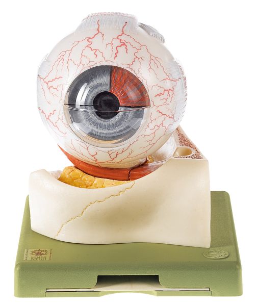 CS 1 Eyeball
 Enlarged approx. 5 times, in SOMSO-Plast®. Resting in the lower bones of the orbit and sectioned horizontally. Separates into 7 parts: sclerotic membrane (2), choroid membrane (2), retina with vitreous humour, lens, bone of the orbit. On...
Price on request
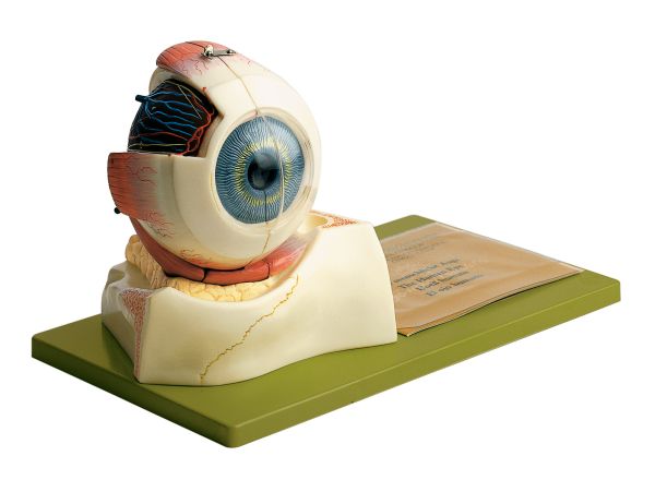 CS 10 Eyeball
 Enlarged approx. 5 times, in SOMSO-Plast®. Resting in the bone of the green base of the orbit. Median section. In the left half, the lens and vitreous humour are fixed. The right half shows the sclerotic membrane partially opened from...
Price on request
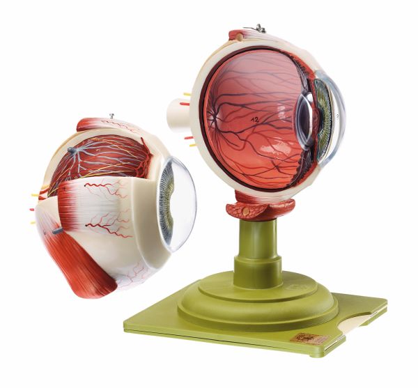 CS 11 Eyeball
 Enlarged approx. 5 times, in SOMSO-Plast®. The eyeball is mounted on the green base. Separates into 2 parts. Median section. In the left half, the lens and vitreous humour are fixed. The right half shows the sclerotic membrane partially...
Price on request
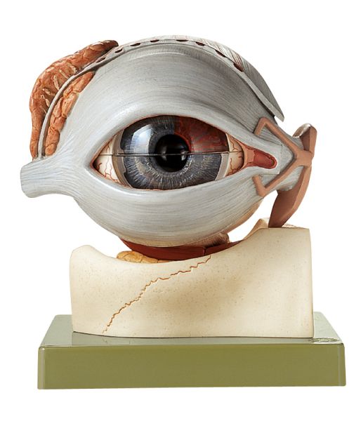 CS 16 Eyeball
 Enlarged approx. 5 times, in SOMSO-Plast®. Resting in the lower bones of the orbit and sectioned horizontally. Sclerotic membrane (2), choroid membrane (2), retina with vitreous humour, lens, bone of the orbit with lacrimal organs and...
Price on request
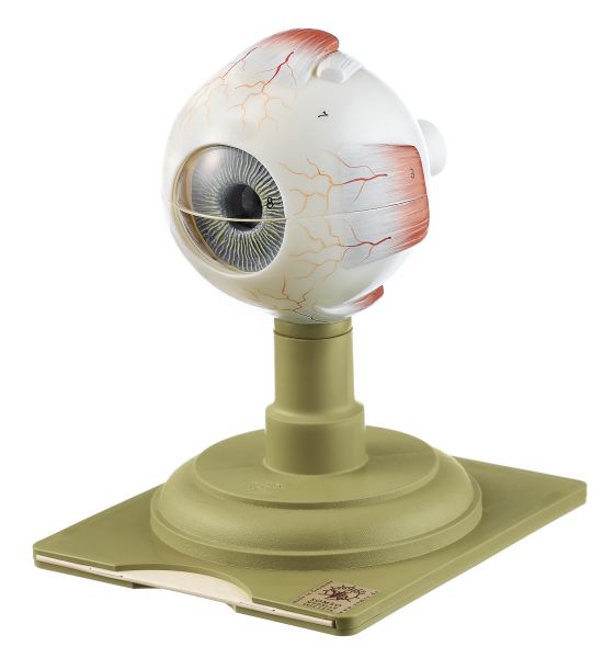 CS 4 Eyeball
 Enlarged approx. 5 times, in SOMSO-Plast®. Sectioned horizontally. Separates into 6 parts: upper half of the sclerotic membrane, choroid membrane (2), Retina with vitreous humour, lens, lower half of the sclerotic membrane. On a stand.
Price on request
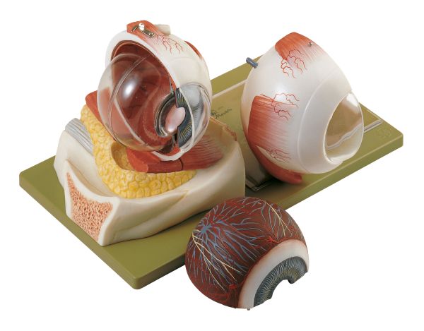 CS 7 Eyeball
 Enlarged approx. 5 times, in SOMSO-Plast®. Resting in the lower bones of the orbit. Separates into 5 parts: Median section of the eyeball (the lens is fixed in the left half), vitreous humour, the right half separates into sclerotic...
Price on request
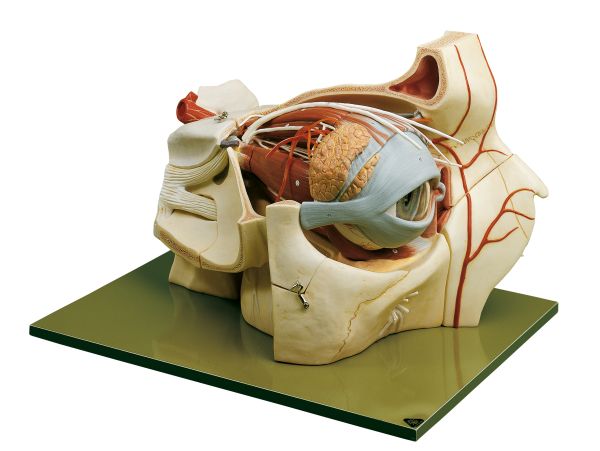 CS 8/1 Topography of the Orbit
 Enlarged approx. 5 times, in SOMSO-Plast®. The orbital process of the frontal bone and the small wing of the sphenoid bone have been removed in order to allow view of the bony orbit. The six muscles of the eye are modelled very clearly...
Price on request
Viewed
